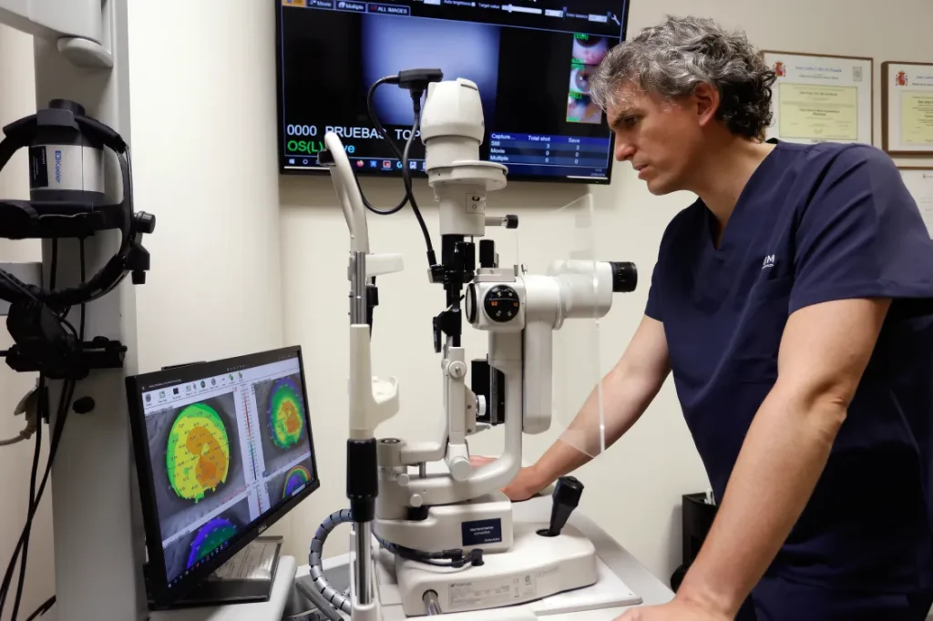
We are leading a study on cell regeneration to strengthen the cornea in patients with advanced keratoconus
A new regenerative medicine technique has shown promising results in the treatment of advanced keratoconus, a disease in which the cornea becomes progressively thinner and changes shape, leading to gradual vision loss.
The research, involving Dr Jorge Alió del Barrio, Medical Director of Vissum Grupo Miranza, evaluated a technique designed to strengthen the cornea by implanting a thin layer of tissue inside the cornea itself. In some patients, this material was also combined with stem cells derived from the patient’s own adipose tissue.
The results have been published in the scientific journal Clinical & Experimental Ophthalmology. The clinical trial was conducted in Spain with patients with advanced keratoconus, who were followed up for one year after surgery.
A thin layer of tissue to strengthen the cornea
Keratoconus causes the cornea to become progressively thinner and develop an irregular shape. As a result, vision can become increasingly blurred and distorted. In the most advanced cases, a corneal transplant may be necessary.
For the trial, the researchers implanted a 120-micron-thick sheet of human corneal stroma into the patients’ corneas. The tissue had previously been decellularised to remove the donor cells while preserving its structural framework.
The participants were divided into three groups: those who received only the corneal tissue sheet; those who received the sheet combined with stem cells obtained from their own adipose tissue; and those who received stem cells alone.
The cornea becomes thicker and patients’ vision improves
One of the study’s main findings was an increase in corneal thickness, which was maintained throughout the one-year follow-up period. Twelve months after surgery, central corneal thickness had increased by an average of 118.4 microns in patients treated with the tissue sheet alone and 121.2 microns in those who received the sheet combined with autologous adipose-derived stem cells.
During the first few weeks, the area where the sheet is implanted may show a slight loss of transparency. This mild opacity gradually disappears, and the corneas regain their transparency within three to six months.
“Cell therapy is going to become a clinical reality; it is now supported by scientific evidence. We have already demonstrated that stem cells can improve established scars. In the future, this will represent a major revolution, although we still need to overcome certain regulatory and standardisation challenges”, explains Dr Alió del Barrio.
The implanted tissue is gradually repopulated
The study also provides insight into what happens inside the cornea after surgery. Over the following months, the researchers observed an increasing number of cells within the implanted sheet. This process also occurred in patients who received the sheet without stem cells, suggesting that cells from the patient’s own cornea can gradually migrate into the new tissue.
Patients who also received stem cells had a greater number of cells within the implanted sheet after one year. The findings provide encouraging evidence regarding the safety and feasibility of this approach over a 12-month period. However, the researchers stress the need for caution and longer-term follow-up to determine whether these results are sustained over time.
Article reviewed by Dr Jorge Alió del Barrio, Medical Director of Vissum Grupo Miranza