 Book an appointment
Eye diseases
Keratoconus
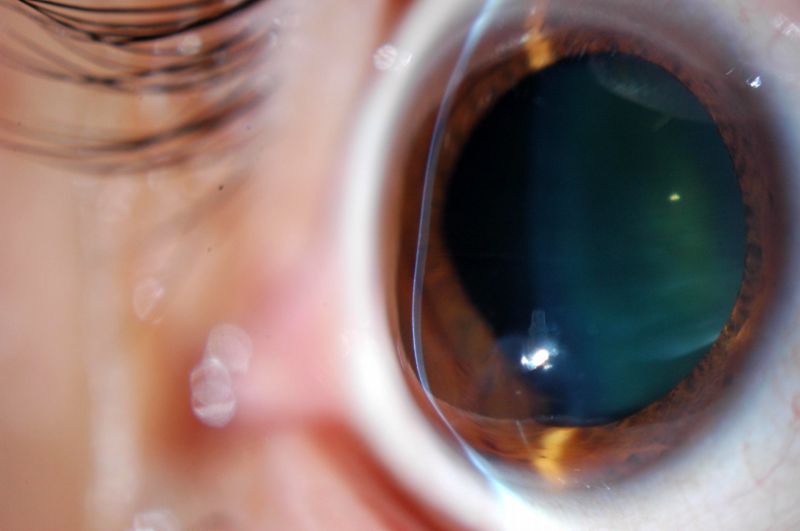
What is keratoconus?
Keratoconus is a degenerative disease caused by the alteration in the collagen fibres that make up the stroma (the thickest part of the cornea). This alteration causes a gradual increase in the curvature of the cornea, which becomes conical (instead of rounded), resulting in gradual vision loss.
It tends to affect both eyes, usually asymmetrically, and is the most common corneal dystrophy, which is present in about 1 in every 2,000 people in Spain.
Symptoms
Causes and risk factors
Diagnosis
Treatment
It is also common for you to experience intolerance to contact lenses and frequent changes in prescription, mainly as regards astigmatism (link), due to the irregularity that is generated on the surface of the cornea. In fact, the appearance or sudden increase of astigmatism – a type of refractive error that generally remains stable and does not evolve – is one of the main signs of suspected keratoconus.



Frequently Asked Questions
Keratoconus is a disease of the cornea that causes progressive thinning and deformation of its shape, making it more curved or cone-shaped. It can cause blurred or distorted vision and frequent changes in prescription. Diagnosis and follow-up make it possible to assess the most appropriate treatment according to how it progresses.
Treatment for keratoconus depends on how advanced it is. In the early stages, glasses or specific contact lenses can be used, while if it is progressing, corneal crosslinking may be indicated to strengthen the structure of the cornea. In advanced cases, surgical techniques or a corneal transplant may be necessary.
Keratoconus can cause distorted perception of images and, in some cases, symptoms that the patient describes as double vision or ghost images, especially with one eye. This is due to the irregularity of the cornea and should be investigated through an eye examination.
Keratoconus can affect both eyes, although not necessarily to the same degree. A person may have more obvious involvement in one eye and milder changes in the other, so follow-up of both eyes is important.
Keratoconus can progress, especially at certain stages of life. Its course is not the same for everyone and it may stabilize in some cases. Regular check-ups make it possible to detect changes in the shape and thickness of the cornea and assess whether intervention is needed to halt its progression.
Keratoconus is detected through an eye examination and tests that analyze the shape and structure of the cornea. Corneal topography and tomography are especially useful for identifying irregularities and monitoring how the disease progresses.
To detect keratoconus, tests such as corneal topography or tomography can be performed, in addition to a complete eye examination. These tests make it possible to study the curvature, thickness and other characteristics of the cornea to determine whether there is an abnormality and to monitor its progression.
How often keratoconus is checked depends on age, the degree of involvement and whether there are signs of progression. The ophthalmologist can set an individual follow-up schedule to monitor changes in the cornea and detect any progression early.
There is currently no treatment that completely eliminates keratoconus, but there are various options to control its progression and improve vision. Treatment depends on the stage of the disease and may include glasses, contact lenses, crosslinking, intrastromal rings or, in advanced cases, a corneal transplant.
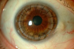
Intracorneal or intrastromal rings are small segments implanted inside the cornea to modify its shape and regularize its surface. They may be indicated in certain patients with keratoconus to improve visual quality and make it easier to fit contact lenses.
The length of a procedure to treat keratoconus depends on the technique used. Crosslinking, intracorneal rings and transplant procedures have different characteristics and surgical times. The ophthalmologist will explain the indicated procedure and its approximate duration before the operation.
In certain cases, surgical techniques involving laser can be used to treat keratoconus, depending on the characteristics of the cornea and the degree of the disease. Not all patients are candidates for the same techniques, so a complete corneal assessment is needed beforehand.
In advanced keratoconus, treatment depends on how much the disease has affected the corneal structure and vision. Techniques such as intracorneal rings may be considered and, when there is significant corneal deterioration that cannot be resolved with other treatments, various types of corneal transplant.
In some patients with keratoconus, special contact lenses are used to improve visual quality when glasses do not provide sufficient vision. There are different lens designs, and fitting must be done on an individual basis to achieve good vision and preserve the health of the ocular surface.
Contact lenses should not be seen as a way to stop keratoconus from progressing. Incorrect fitting or improper use can cause ocular surface problems, so it is important that lenses are prescribed and monitored by specialist professionals.
Keratoconus can be treated and controlled with various options, although the approach depends on how advanced it is. Crosslinking can be used to halt progression in selected patients, while contact lenses, intracorneal rings or other techniques can help improve vision. In advanced cases, a corneal transplant may be necessary.
If keratoconus progresses unchecked, the cornea can become progressively deformed and thinner, causing a deterioration in visual quality. In advanced stages, corneal scarring or opacities may appear and it may be harder to achieve good vision with glasses or contact lenses. Follow-up makes it possible to detect progression and consider treatment when necessary.
Keratoconus can have a family component, and there are genetic factors that can increase the predisposition to develop it. For this reason, when a family member has keratoconus, eye check-ups may be advisable, especially during childhood and adolescence.
Keratoconus usually begins during adolescence or in young adults, although it can be detected at different ages. Its progression varies and, in some cases, it may progress more quickly during the first few years. This is why detection and follow-up are especially important in young patients.
Frequent or vigorous eye rubbing has been linked to the progression of keratoconus and can damage the ocular surface. People with this disease should avoid this habit and see their ophthalmologist if they have frequent itching or discomfort so that the cause can be identified and treated.
Keratoconus can cause a significant loss of visual quality and acuity if it progresses, but there are treatments aimed at controlling its progression and improving vision. In advanced stages, surgical procedures can be used, including corneal transplant when indicated.
There is no guaranteed way to prevent keratoconus, but it is important to avoid rubbing your eyes and to have eye check-ups when there are risk factors or changes in vision. Detecting the disease and monitoring its possible progression makes it possible to consider treatment early.
Yes. Corneal irregularity can make it difficult to see clearly in low light, as well as causing halos, glare or distorted images. These symptoms can be especially troublesome when driving at night and should be assessed through an eye examination.
Surgery is not necessary for all patients with keratoconus. The decision depends on how the disease progresses, visual quality, the shape and thickness of the cornea and the response to other treatment options. A complete corneal assessment makes it possible to determine whether surgery is indicated and which technique may be most appropriate.
There is no single treatment that is best for all patients with keratoconus. Depending on the stage of the disease, glasses, contact lenses, crosslinking, intracorneal rings or other surgical techniques can be used. Treatment should be decided after studying the cornea and assessing whether it is progressing.
Keratoconus may rule out certain laser refractive surgery techniques, as the stability and structure of the cornea need to be assessed beforehand. Before considering surgery to correct myopia, a complete corneal assessment is essential to determine which options are safe and appropriate.