Retina: Understanding Its Diseases and Discovering the Latest Treatments at Miranza
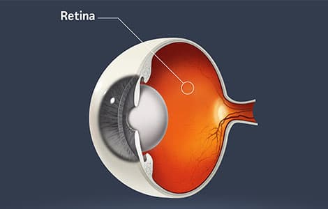
The retina, located at the back of the eyeball, is a thin layer of light-sensitive tissue that plays a crucial role in our ability to see. Through it, the light entering the eye is converted into electrical signals, which travel through the optic nerve to the brain, where they are interpreted as images. Its proper functioning is essential for clear and detailed vision, and its complexity makes it one of the most delicate and fascinating areas of the visual system.
What the retina is like, a key part of the eye
The retina lines the inside of the eye like a highly specialized biological wallpaper. It is composed of several layers of nerve cells, including cones and rods, which allow the perception of color, light, and movement. These cells are constantly active, communicating with each other and with the brain through electrical signals. The retina is nourished by blood vessels and bathed by the vitreous humor, a gelatinous substance that occupies the center of the eye. This entire system is carefully organized to maintain precise and continuous vision.
How the retina works: the vision process
When we open our eyes and observe our surroundings, what actually happens is that light passes through various ocular structures until it reaches the retina. There, the cones and rods react to light stimuli, generating electrical impulses. These signals are processed by the inner layers of the retina and transmitted through the optic nerve. It is a sophisticated and continuous process that allows us to distinguish shapes, colors, contrasts, and even orient ourselves in darkness. Any disruption in this mechanism can have significant consequences for the visual field.
Main Retinal Diseases
Retinal diseases are a common cause of vision loss, especially in older adults or individuals with certain medical conditions. Although each disease has its own characteristics, they all share a potential impact on visual quality, making early detection and proper treatment essential.
Age-Related Macular Degeneration (AMD)
AMD affects the macula, the central area of the retina responsible for detailed vision. It can develop in either a dry or wet form and is one of the leading causes of visual deterioration in older adults.
Diabetic Retinopathy
Diabetes can damage the blood vessels in the retina, causing microhemorrhages, exudates, and edema. Diabeticretinopathy often progresses silently until advanced stages. Therefore, regular check-ups are essential for those with this systemic disease.
Retinal Detachment
When the retina separates from the inner wall of the eye, it creates a medical emergency. Retinal detachment disrupts the visual process and can lead to permanent vision loss if not treated promptly.
Macular hole
A macular hole is a condition that causes a small opening in the macula, the central part of the retina responsible for detailed vision. This lesion can make tasks like reading, driving, or recognizing faces difficult. Although its progression can vary, it usually requires surgical intervention through a vitrectomy. You can find more information about macular hole and its treatment at Miranza.
Epiretinal membrane
An epiretinal membrane, also known as macular pucker, is a thin layer of fibrous tissue that forms over the macula. This membrane can contract and wrinkle the retina, leading to distorted or blurry vision. While its progression can be slow in some cases, not requiring immediate intervention, others may benefit from surgery to restore the macular anatomy and improve visual quality.
Retinal vascular occlusions
When a retinal vein or artery gets blocked, blood circulation is interrupted. This can lead to inflammation or sudden vision loss. Quick intervention is crucial to preserve the affected visual field.
Central serous chorioretinopathy
This condition involves a buildup of fluid under the retina, causing blurry vision and central distortion. While it often resolves on its own, some patients need specialized monitoring to prevent recurrences.
Retinitis Pigmentosa and Other Hereditary Dystrophies
Retinitis pigmentosa is a hereditary disease that progressively degenerates the rods and cones. It often begins with night and peripheral vision loss, and can evolve into a severe reduction of the visual field. There are other retinal dystrophies with different mechanisms and prognoses, so a genetic and functional evaluation is fundamental.
Signs indicating a possible retinal problem
Detecting a retinal disease early is key to preserving vision. Some common manifestations are:
- Perception of flashes or lights (photopsias)
- Sudden appearance of floaters in the visual field
- Distortion of straight lines or the loss of central or peripheral vision
These symptoms can develop progressively or suddenly, so it’s advisable to see a specialized ophthalmologist for any visual change, even if it seems mild.

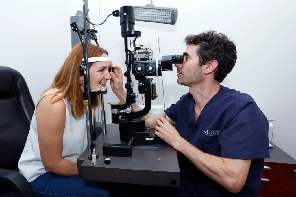
Diagnostic methods for retinal problems at Miranza
Diagnosing retinal diseases requires precision and advanced technology. At Miranza, we use various tests to observe the condition of the fundus and the inner layers of the retina:
- Retinography allows us to obtain high-resolution images.
- Angiography evaluates the circulation of retinal blood vessels.
- OCT and its variant, Angio-OCT, offer a detailed scan of retinal structures without contact or pain.
These tools, along with a complete clinical examination, allow us to detect alterations even before they affect vision. You can learn more about these procedures in our article on eye and fundus examinations.
Advanced treatments for retinal diseases
Treatment for a retinal condition is always decided after a personalized medical evaluation, considering the type of disease, its progression, and the patient’s visual status. Intravitreal injections are one of the most commonly used treatments today, especially for macular degeneration or macular edema. We also have laser techniques, like photocoagulation, which are useful for treating abnormal vessels or preventing hemorrhages. In more complex situations, such as a retinal detachment or a vitreous hemorrhage, surgical intervention via vitrectomy may be necessary. This technique allows access to the inside of the eye to repair the retina.
Retina prevention and care
At Miranza, different therapeutic options are applied depending on the patient’s condition and visual status. One of the most common treatments is intravitreal injections, used to administer medications directly into the eye and treat conditions like macular degeneration or diabetic retinopathy.
In other cases, laser application via photocoagulation may be necessary. This technique seals off abnormal vessels or prevents complications in the early stages of some retinal diseases.
Another fundamental surgical technique is vitrectomy, indicated for treating vitreous hemorrhages, macular holes, or epiretinal membranes. It allows access to the inside of the eye to remove the vitreous humor and resolve tractions affecting the retina.
Why choose Miranza for your retina care
At Miranza, we approach retinal health with a comprehensive focus, combining clinical experience, high medical specialization, and cutting-edge diagnostic and therapeutic technology. Our ophthalmology teams work in coordination to offer personalized care, based on scientific evidence and knowledge accumulated over decades of dedication to ophthalmology. Our commitment is to support each patient from prevention to treatment, with the rigor and closeness they deserve.