Diagnostic tests
Optical Coherence Tomography (OCT)
What is OCT?
OCT or Optical Coherence Tomography is a diagnostic imaging technique that, in recent years, has become essential in ophthalmology consultations. With micrometric precision, it allows us to visualise different eye structures in 3D, performing an automatic “scan” that provides us with highly relevant information for the detection and monitoring of many eye diseases.
Its main advantage is that it is a non-invasive technology with a much higher resolution than other techniques used in medicine that also make optical “cuts” across tissues (such as CT or MRI).
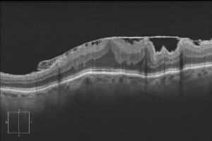
To do so, an OCT works based on the emission of infrared light that, when reflected on the eye structures, shows us the layers or sections of different parts of the eye, hence detecting very subtle alterations, practically on a cellular level.
Depending on the structures it is focused on, we can distinguish two types of OCT:
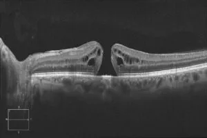
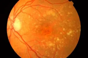
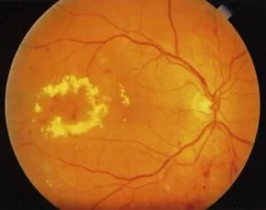
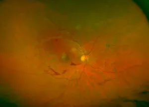
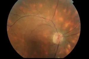
- Optical Coherence Tomography of the posterior pole: visualisation of the retina and, with the latest models, even of the layer behind, i.e. the choroid.
- Optical Coherence Tomography of the anterior pole: visualisation of the cornea, the anterior chamber of the eye, the iris and the lens. This can be done with posterior OCT equipment to which specific software or lenses are adapted, or with new equipment specifically for anterior OCT.
What do we use OCT for?
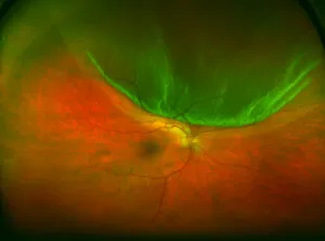
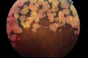
Some of the main pathologies for which we use posterior OCT.
In turn, anterior OCT is also indicated for a wide variety of diseases, as well as before and after certain ophthalmic surgeries.
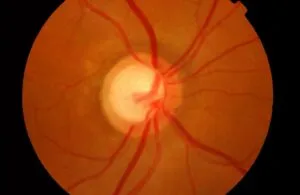
In the case of glaucoma, both posterior OCT, as it allows us to study the head of the optic nerve damaged by the disease, and anterior OCT, which provides us with data on the eye structures of the anterior chamber of the eye on which the treatments of this pathology act, are useful.

How do we perform OCT?
- It is a very comfortable and quick test, which takes approximately 5 to 10 minutes.
- It is painless, as it does not involve contact with the eye and avoids having to use anaesthetic drops.
- It can be performed without dilating the eye (in some cases, we must necessarily do it without dilating the pupil).
- We perform it in our consulting rooms, which are equipped with the latest diagnostic technology at the Miranza clinics.
- We get the results immediately to complete your diagnosis.